Answered Sep 24, 2018. Hyper means more and hypo means less, so hypodense meansless dense than average. CT scans are literally just shades of grey based on how dense the tissue is when an x-ray tries to travel through it. Air is least dense and is black. Fat and water are hypodense and dark grey..
Just so, what is hypodense in CT scan?
A hypodensity usually means we have a decreased water content or water movement. A hypodensity usually means we have a decreased water content or water movement. That can be due to different things, but usually this is something you would see during ischemic stroke injury.
Furthermore, what does Hypodense liver mean? Liver lesions are groups of abnormal cells in your liver. Your doctor may call them a mass or a tumor. Noncancerous, or benign, liver lesions are common. They don't spread to other areas of your body and don't usually cause any health issues. But some liver lesions form as a result of cancer.
Simply so, what is Hypodense?
hypodensity. Noun. (plural hypodensities) (medicine) An area of an X-ray image that is less dense than normal, or than the surrounding areas.
Why is blood Hyperdense on CT?
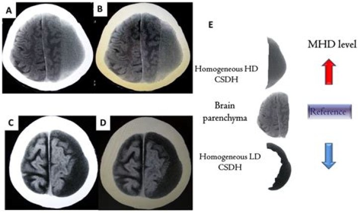
Hyperdensity at CT was due to the high hemoglobin content of retracted clot or sedimented blood. The various patterns seen can be related to sequential changes occurring in blood following hemorrhage. Relative hyperdensity and its variations seen on precontrast scans are useful diagnostic signs of recent hemorrhage.
Related Question Answers
What does Isodense mean?
isodense. Adjective. (not comparable) (sciences, especially biochemistry) Evenly or uniformly dense; of the same density (as an adjacent object, tissue, etc).What does hypodense mass mean?
On ultrasound, the fatty areas are hyperechoic and ill defined. On CT, the lesions are hypodense, without mass effect on adjacent structures, and often have normal sized vessels passing through them. On MRI, lesions are hyperintense on T1 weighted images and disappear with fat suppressed images.What causes hypodense lesions in the liver?
Colon, lung, breast, and gastric cancers are the most common causes of hypovascular liver metastases. If the lesions do not show this appearance, small hypodense metastases may be difficult to differentiate from a host of benign hepatic lesions.What does white mean on CT?
The white area signals dense tissues like bone, the gray area represents soft tissues and fluids, and the dark gray and black area shows air and fat.What color is blood on a CT scan?
Blood. Look for the presence of blood. Clues to the origin of the haemorrhage, its duration and the cause of the insult may be indicated by its position and spread. Acute haemorrhage absorbs X-rays and appears hyperdense (white) on CT scans.What does Hounsfield mean?
Definition/Introduction The Hounsfield unit (HU) is a relative quantitative measurement of radio density used by radiologists in the interpretation of computed tomography (CT) images. The absorption/attenuation coefficient of radiation within a tissue is used during CT reconstruction to produce a grayscale image.What is bright on CT?
Tissues like air and water have little attenuation and are displayed as low densities (dark), whereas bone has high attenuation and is displayed as high density (bright) on CT. Among pathologic conditions, high density lesions are often seen with freshly clotted blood, hyperemia and with the use of contrast.Are hypodense lesions cancerous?
Metastases are the most common malignant liver tumors, and occur 20 times more frequently than primary hepatic neoplasms[2]. If the lesions do not show this appearance, small hypodense metastases may be difficult to differentiate from a host of benign hepatic lesions.What does Hypoenhancing mean?
Definition: Enhancement that unequivocally is less than that of liver. If there is equivocal hypoenhancement: Characterize as isoenhancement.What does focal Hypodensity mean?
Focal= small areas. Hypodensities= Which are less dense than surrounding tissues. may represent. perivascular=area surrounding blood vessels.What causes Hypodensity in the brain?
Brain lesions can be caused by injury, infection, exposure to certain chemicals, problems with the immune system, and more. Typically, their cause is unknown.What is kidney Hypodensity?
Small hypodense renal lesions with a round shape are frequently detected on CT scans of the upper abdomen after contrast medium administration. In nearly all cases these round hypodensities are simple small cysts with no clinical significance.Can you die from white matter disease?
White matter disease has been implicated in tissue and clinical outcomes of patients with acute ischemic stroke, and data link white matter disease burden measured semiquantitatively and functional dependence or death in patients with spontaneous primary brain hemorrhage, according to the investigators.What is subcortical Hypodensity?
Background. Subcortical hypodensities of presumed vascular etiology (SHPVO) are radiological findings defined as rounded ill-defined areas of decreased attenuation on CT with increased signal on T2-weighted MR sequences such as fluid-attenuated inversion recovery [1, 2].How do you describe a CT scan?
A computerized tomography (CT) scan combines a series of X-ray images taken from different angles around your body and uses computer processing to create cross-sectional images (slices) of the bones, blood vessels and soft tissues inside your body. CT scan images provide more-detailed information than plain X-rays do.What does Hypodense mean on CT?
Hyper means more and hypo means less, so hypodense meansless dense than average. CT scans are literally just shades of grey based on how dense the tissue is when an x-ray tries to travel through it. Air is least dense and is black. Fat and water are hypodense and dark grey. Bone is hyperdense and bright white.What is liver parenchyma?
The liver parenchyma is the functional tissue of the organ made up of around 80% of the liver volume as hepatocytes. The other main type of liver cells are non-parenchymal. Non-parenchymal cells constitute 40% of the total number of liver cells but only 6.5% of its volume.What is the first sign of liver problems?
The first symptoms of liver failure are often nausea, loss of appetite, fatigue, and diarrhea. Because these symptoms can have any number of causes, it may be hard to tell that the liver is failing.What is the first sign of liver cancer?
The signs and symptoms of liver cancer are most often the result of liver damage and may include yellowing of the skin (jaundice), right-sided abdominal or shoulder blade pain, or a lump in the right upper abdomen. However, many of the warning signs are non-specific, such as weight loss and fatigue.